
Expert Retina Care in Kurukshetra — Specialist Diagnosis & Treatment
The retina is the most delicate and vital part of your eye. When something goes wrong with it — diabetic damage, a tear, macular degeneration — fast, expert care makes the difference between keeping and losing your sight. Dr. Amit Sobti, MS Ophthalmology AIIMS, provides comprehensive retinal services at Sobti Nursing Home, Kurukshetra.
Seek Urgent Care if You Notice These
These symptoms can indicate a retinal emergency — do not wait
Lightning-like flashes in your vision — especially new or worsening flashes
New floaters — especially many at once — can signal a retinal tear
A dark shadow spreading across your visual field — this is a retinal emergency
Straight lines appearing wavy or bent — a sign of macular disease
If you notice any of these symptoms, call us immediately on 98176-33316. A retinal detachment treated within hours has a much better outcome than one treated days later.
Retinal Conditions Treated at Sobti Nursing Home
From diabetic retinopathy to macular degeneration — comprehensive retinal care under one roof.
Diabetic Retinopathy
The leading cause of preventable blindness in India. Diabetes damages the tiny blood vessels in the retina — often silently until significant vision is lost. Annual screening is essential for all diabetic patients.
Retinal Detachment
The retina peels away from the back of the eye. A time-critical emergency — if treated promptly, vision can be preserved. Symptoms: sudden flashes, floaters, or a curtain in vision. Call immediately.
Retinal Tears & Holes
A tear in the retina can lead to detachment if not treated promptly. Laser photocoagulation seals the tear and prevents progression. Best treated within days of the first symptoms.
Macular Degeneration (ARMD)
Age-related macular degeneration affects central vision — the sharp, detailed vision used for reading, recognising faces, and driving. Both dry and wet forms are managed at our clinic.
Diabetic Macular Oedema
Fluid accumulation in the macula — the central retina — causing blurred or distorted central vision in diabetic patients. Treated with anti-VEGF injections and laser with excellent results.
Retinal Vein Occlusion
Blockage of the retinal vein causes haemorrhage and swelling inside the eye, leading to sudden vision loss or blurring. Early anti-VEGF treatment significantly improves visual outcomes.
Vitreous Haemorrhage
Bleeding into the vitreous gel of the eye — causing sudden vision loss or a shower of floaters. Often secondary to diabetic retinopathy or retinal tears. Requires urgent assessment.
Epiretinal Membrane
A thin membrane growing on the macula causes distortion and blurring of central vision. Managed with observation initially, and surgery when vision is significantly affected.
Central Serous Retinopathy
Fluid accumulates under the macula — common in young to middle-aged men under stress. Causes a dark spot or distortion in central vision. Usually self-limiting but needs monitoring.
Why the Retina Matters So Much
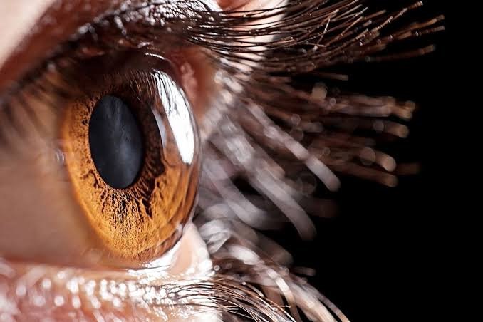
The retina is a thin layer of light-sensitive tissue lining the back of the eye. It works like the film in a camera — capturing the visual image and converting it into electrical signals that travel via the optic nerve to the brain, where the image is processed.
At the centre of the retina is the macula — a tiny but critically important area responsible for all sharp, detailed, central vision. Reading, recognising faces, watching TV, driving — all of this depends on the macula working perfectly.
Unlike many other tissues in the body, the retina has very limited ability to repair itself. Damage to retinal cells is often permanent. This is why early detection and prompt treatment are not optional — they are essential.
A 5mm area at the retina's centre. Responsible for all fine detail vision — reading, faces, driving. The target of ARMD and diabetic macular oedema.
Responsible for movement and navigation. Affected by retinal detachments and peripheral retinal tears — which can extend inward if not treated.
Supply oxygen and nutrients to the retina. Damaged in diabetes and hypertension — leading to bleeding, leakage, and eventually vision loss.
Advanced Retina Imaging & Diagnostics
Accurate diagnosis requires high-quality imaging. We use modern equipment to see what the naked eye cannot.
OCT — Optical Coherence Tomography
Cross-sectional imaging of the retinal layers at microscopic resolution. Essential for diagnosing and monitoring macular disease, DME, and ARMD. Non-invasive, takes seconds.
Fundus Photography
High-resolution photographs of the retina, optic disc, and blood vessels. Provides a permanent record for monitoring disease progression over time.
Fluorescein Angiography (FFA)
A dye is injected into the arm vein and photographs track its passage through the retinal vessels — revealing leakage, blockages, and areas of abnormal blood vessel growth.
Indirect Ophthalmoscopy
A detailed clinical examination of the entire retina after dilating drops — essential for peripheral retinal assessment, tears, and detachment evaluation.
How We Treat Retinal Conditions
Treatment depends on the specific condition, its severity, and how quickly it is caught. Dr. Sobti will explain all options clearly.
Laser Photocoagulation
A focused laser seals leaking blood vessels, destroys abnormal vessels, or creates a barrier around retinal tears to prevent detachment. Painless, performed in clinic.
Anti-VEGF Injections
Intravitreal injections of anti-VEGF medicine reduce abnormal vessel growth and macular swelling. Highly effective with minimal discomfort. Administered under local anaesthetic drops.
Intravitreal Steroids
Steroid injections into the vitreous reduce macular inflammation and oedema. Used in certain cases of macular oedema where anti-VEGF therapy alone is insufficient.
Vitreoretinal Surgery
For retinal detachments, vitreous haemorrhage, and epiretinal membranes requiring surgical intervention. Dr. Sobti will advise if surgical referral is needed for complex cases.
Diabetic Retinopathy — The Most Important Thing Every Diabetic Patient Must Know
Diabetic retinopathy is the leading cause of preventable blindness in working-age adults in India. It affects 1 in 3 people who have had diabetes for more than 20 years — and causes no symptoms until it is already advanced.
The most important fact: you can have significant diabetic retinopathy with perfect vision. By the time your sight deteriorates, the window for the most effective treatment may already be closing.
Annual retinal screening is not optional for diabetic patients — it is as essential as checking your blood sugar.
Tiny swellings in retinal blood vessels. No vision symptoms. Managed with good diabetes control and annual monitoring.
More blood vessels are blocked. Laser treatment may be needed. Tighter diabetic control is essential to slow progression.
Abnormal new vessels grow on the retina — fragile, likely to bleed. Laser and injections are urgently needed to prevent severe vision loss.
Diabetic Retinopathy — Key Facts

Retina Care by an AIIMS-Trained Eye Specialist
Retinal conditions require specialist expertise, advanced imaging, and prompt decision-making. Dr. Amit Sobti brings AIIMS-level training to Kurukshetra — so you don't have to travel to a city for expert retinal care.
Comprehensive retinal training at India's premier ophthalmology programme. You get the expertise of a city specialist without leaving Kurukshetra.
Microstructural imaging of the retina for precise diagnosis. Essential for detecting and monitoring macular disease accurately.
Retinal emergencies cannot wait. We prioritise urgent cases — call 98176-33316 if you have sudden vision symptoms and we will see you the same day.
Retinal treatment including injections and laser is covered under ECHS for eligible patients. We handle all the paperwork.
Retina — Common Questions
Don't Wait With Your Retinal Health
Retinal conditions worsen quickly and the window for effective treatment can be narrow. Book your consultation today — same-day appointments available for urgent cases.